The Rise of Digital Radiography in Veterinary Medicine
Advanced imaging modalities like digital radiography are quickly gaining popularity in veterinary medicine. In fact, studies have shown that the global veterinary diagnostic imaging market, which amounted to USD 711.45 million in 2018, is expected to witness a compound growth rate of 6.45% from 2019-2024.
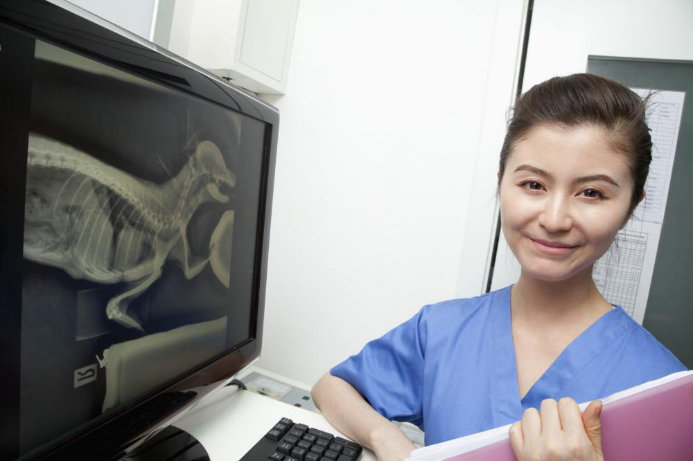
X-ray systems use gamma radiation at a low dose rate to create a two-dimensional, detailed image of the pet’s abdominal cavity, chest and bony structures.
Although the technology associated with X-ray generation has remained relatively unchanged since its discovery, the radiograph production process has evolved significantly over the years. The introduction of digital radiography systems, in particular, has revolutionized the efficiency of radiograph dissemination and production.
How Direct Digital Radiography Helps Improve Pet Patient Care
Direct digital radiography (DR) is a method of digital radiography that replaces the conventional radiographic cassette and film with a flat panel detector that acts as the digital imaging sensor. The detector connected to a computer converts the X-ray beams into electrical pulses and then into an image that can be viewed immediately after exposure. In veterinary clinics, the flat panel detector may be affixed to an X-ray table or attached to a DR computer by a wire cable.
Digital radiography has become the gold standard in veterinary practices, even since its introduction over 15 years ago. What started out as a costly investment when first launched has now become mainstream and affordable.
Let’s look at how the digital diagnostic imaging system supports better patient outcomes and expands consultation capabilities in veterinary clinics:
Fast and Easy Way to Examine Pets
Conventional film radiography takes time to process since each image can only be assessed after it comes out of the processor and is placed in a lightbox. Digital radiography, on the other hand, can be viewed immediately after exposure.
In veterinary medicine, this advantage is enormous. It allows veterinarians to adjust the pet’s position and retake the image without ever moving the animal off the table.
Reduced Radiation Exposure
Since digital radiography systems offer a wide viewing area and greater exposure latitude, the number of retakes required to produce a radiograph significantly decreases. And fewer retakes mean decreased radiation exposure to the patients and staff.
Moreover, digital sensors require less radiation per exposure to produce high-quality radiograph. Needless to say, clients appreciate the lowered radiation doses for their pets.
Exceptional Image Quality
It isn’t easy to get a clear read from traditional film radiography due to issues arising from its technique-dependent nature. Moreover, zooming in or changing the brightness of the image is also impossible with film-based systems.
By switching to DR, veterinarians can reduce the number of poor-quality radiographs that result from exposure errors. Plus, the image processing algorithms used in digital X-ray systems are much more forgiving; the software allows for adjustment of contrast, brightness, pan and zoom on a single exposure. As long as the pet patient is correctly positioned, almost everything else can be fixed with the software. Veterinarians can expect a balanced presentation of overlapping soft tissue and bone structures.
Upgrading to digital X-ray solutions is a necessary investment if veterinarians wish to improve efficiency and enhance diagnostics. DR also offers improved imaging latitude, access to teleradiology and unparalleled ease-of-use.
Therefore, it doesn’t come as surprise that digital radiography, today, is galloping full-tilt into mainstream use.
By investing in state-of-the-art digital imaging solutions, like ExamVue’s Digital X-Ray systems and DR acquisition software, DVMs can enjoy high-quality images and rapid diagnosis. Additionally, the images are stored using a DICOM standard that allows veterinarians to transmit, store and retrieve medical imaging information.
What’s more, our high-quality DR flat panel detectors focus on convenience and durability. They work together with the imaging software to offer a vast range of image manipulation options.
ExamVue also provides digital radiology equipment to podiatrists, orthopaedists, chiropractors, and other medical specialties. Get in touch with us today for more information on our products.


 Reduced Radiation Exposure