Digital images are essentially images in the form of computer files. Radiography images are large in terms of size with high spatial and contrast resolution. All radiographic images must be DICOM, which is a standard for Digital Imaging and Communications in Medicine, for medical use. DICOM files combine images, patient data, and image descriptions, such as anatomy, time, etc. DICOM files limit what you can do with them. This includes: producing, viewing, printing, saving, sending, and several others.
Some sources of digital images include the scanning of x-ray film, scanning of Computed Radiography (CR) plates, Flat Panel Detectors (FPD), and Charge Coupled Detectors (CCD). CR was commercialized in 1983, making it the oldest digital technology. This technology uses a cassette and reader. The cassette contains a Photostimulable Storage Phosphor (PSP) which is intended to be scanned, erased, and reused. Cassette sized and use is similar to film x-ray.
CCD is a digital camera technology in which the screen turns x-rays into light. Camera lenses focus light onto a small chip and light is changed into electrical current.
CR versus CCD:
• Easier retrofit
• Cassettes enable off-table shots
• Slightly less expensive in short term
• Least change to workflow (could perceived as good or bad)
On the other hand, when compared to CR, CCD has much faster image acquisition, longer life, no scanner, no cassettes to damage, and no cleaning required. CCD is also repairable.
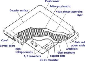
Flat Panel Detectors (FPD) are very common in digital radiography (DR). They place Thin Film Transistors (TFT) directly in the path of x-rays. These panels almost always utilize a scintillator screen, which turns x-rays into visible light. Scintillators are produced from Gadolinium Oxide (“Gadox”) or Cesium Iodide (CsI), which is preferable due to its higher efficiency. CsI, however, does come at a premium.
You can learn more about ExamVue flat panel detectors here >>


